Case Of the Week (COW) 06 Oct 2013
Answer:
Left Subclavian artery steal.
Findings:
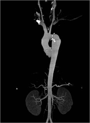
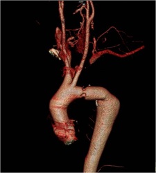
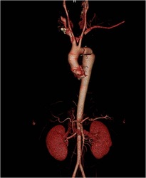
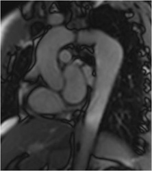
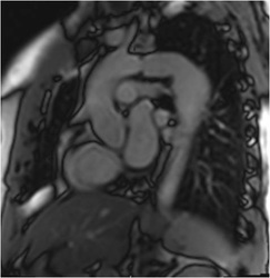
Phase Contrast Flow Velocity Imaging There is left Subclavian artery steal with caudo cranial flow in right vertebral artery(white) and craniocaudal flow in left vertebral artery(black). The velocity versus time curve shows similar findings. CT Angiogram demonstrates the absent flow in proximal left subclavian artery with reconstituted flow via collaterals and vertebral artery. Mild focal luminal narrowing noted at site of graft.
Discussion:
Common causes of subclavian steal phenomenon are stenosis /occlusion of subclavian artery or sometimes Arteriovenous fistulas. This person had a previous coarctation of Aorta with left subclavian origin aneurysm for which Hemashield graft and ligation of Proximal subclavian artery was done. This was the unusual cause of the reversal of flow in left Vertebral artery.
Contributed By:
Dr. Babu Peter MD, DNB
Associate Professor, Barnard Institute of Radiology
Senior Consultant Radiologist, Aarthi Scans, Chennai
